Can Dental X-Rays Detect Cavities Between Teeth? What Bitewing Images Reveal
The correct quiz answer is A: Cavities between teeth. Dental X-rays can detect cavities between neighboring teeth that may be difficult or impossible to see during a visual examination. Bitewing dental X-rays are particularly useful because they show the contacting surfaces of neighboring back teeth, where decay can develop unnoticed. However, very early cavities may not appear on an X-ray, so dentists combine imaging with clinical examination and individual cavity risk assessment.
Key Takeaways:
Bitewing X-rays help detect hidden decay between neighboring teeth.
Very early enamel changes may not be visible because sufficient mineral loss must occur before a radiographic difference can be detected.
A suspicious X-ray finding does not automatically require a filling. Some early, noncavitated lesions may be managed with preventive care and monitoring.
Dental imaging frequency should depend on cavity risk, symptoms, clinical findings, and previous images rather than a universal schedule.
Modern dental X-rays involve relatively low radiation exposure, and imaging should be selected according to clinical need.
Dental X-rays do not show ordinary tooth colors or surface stains the way photographs do.
This guide explains how bitewing dental X-rays work, which problems they can reveal, how findings are interpreted, and when dental imaging may be recommended.
Medical information notice: This article provides general educational information and does not replace individualized examination, diagnosis, or treatment by a qualified dental professional.
Quiz Answer: Which Problem Can Dental X-Rays Reveal That May Be Difficult to See?
Cavities between teeth is the correct answer. Dental X-rays can reveal mineral-density changes on tooth surfaces that are difficult to examine directly, particularly where neighboring back teeth contact each other.
Bitewing radiographs help dentists evaluate these hidden surfaces. Areas affected by sufficient mineral loss may appear darker than healthy enamel or dentin, providing evidence of possible tooth decay.
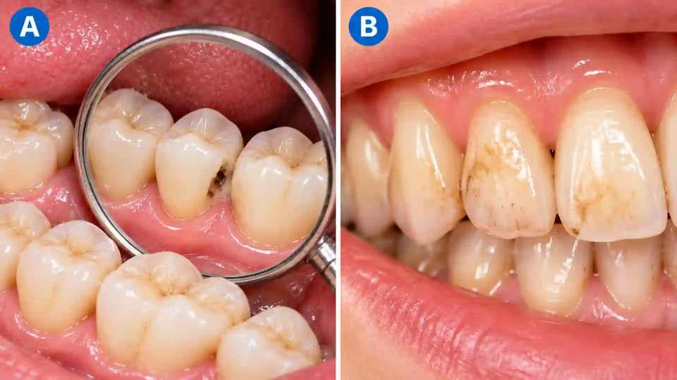
Surface stains are not the correct answer. Dental X-rays do not record natural tooth colors like ordinary photographs. Brown or yellow stains on enamel may be visible during an oral examination without producing a detectable change on an X-ray.
A dental X-ray is one part of the diagnostic process. Dentists also consider clinical findings, cavity risk, symptoms, and previous images before recommending treatment.
How Do Bitewing Dental X-Rays Detect Cavities Between Teeth?
Bitewing dental X-rays help dentists identify tooth decay by showing differences in the density of tooth structures. Areas that have lost sufficient minerals may appear darker than the surrounding healthy enamel or dentin.
This makes bitewing imaging particularly useful for identifying interproximal caries, the clinical term for decay developing on surfaces where neighboring teeth contact each other.
The contacting surfaces of neighboring teeth are often hidden from direct view. A dentist can inspect visible and chewing surfaces, but tightly contacting teeth can conceal changes occurring between them.
Food debris and dental plaque may collect in these spaces. When plaque bacteria metabolize sugars and other fermentable carbohydrates, they produce acids that can gradually remove minerals from enamel.
Decay can progress without obvious pain or a visible hole. Someone may therefore have cavities between teeth even when their teeth appear healthy in a mirror.
How Does Tooth Decay Appear on a Dental X-Ray?
Dental X-rays pass through oral tissues and are recorded by a digital sensor or another imaging receptor.
Dense structures absorb more X-ray radiation and generally appear lighter on the image. Areas with reduced mineral density allow more radiation to pass through and may appear darker.
A darker area on a radiograph is called a radiolucency. When its location and pattern are consistent with tooth decay, the dentist may interpret it as a suspected carious lesion.
However, not every radiolucency represents decay. Normal anatomy, imaging artifacts, and other conditions can produce similar appearances.
For this reason, a dentist must interpret the image alongside the clinical examination. Identifying a dark area on an X-ray is not enough to establish a diagnosis or determine whether a filling is needed.
What Can Bitewing Dental X-Rays Reveal?
Bitewing X-rays can reveal signs of tooth decay, the approximate radiographic depth of some lesions, existing restorations, suspicious changes around certain fillings, and portions of the bone supporting the teeth.
Cavities between teeth: Bitewings may show darker areas near the contacting surfaces of neighboring teeth. Very early lesions may not be visible.
Enamel decay: Mineral loss may appear within the outer tooth layer. Radiographic appearance does not always indicate the exact extent of actual tissue damage.
Dentin decay: A suspicious area may extend into the less-mineralized tissue beneath enamel. Treatment depends on clinical findings as well as apparent depth.
Decay near existing fillings: Changes around restoration margins may suggest recurrent decay, although imaging artifacts and restoration materials can complicate interpretation.
Supporting bone levels: Bitewings can show portions of the bone between neighboring teeth, but one image alone cannot diagnose every periodontal condition.
Existing restorations: Fillings, crowns, and visible restoration margins can be examined, although some materials obscure underlying structures.
The main benefit is additional information that complements rather than replaces the clinical examination.
Can Dental X-Rays Show Surface Stains or Tooth Color?
Dental X-rays are not ordinary color photographs. They display differences in X-ray absorption by tissues and materials rather than the natural colors of teeth.
A brown stain on enamel may be readily visible during a clinical examination but may not produce any detectable change on a radiograph.
Likewise, a darker area on an X-ray does not necessarily correspond to a visible brown or black spot on the tooth.
Surface staining, enamel demineralization, and physical cavitation are different findings. Dentists use visual examination, radiographic information, clinical history, and other appropriate methods to distinguish them.
This distinction explains why cavities between teeth are the correct quiz answer rather than surface stains.
Which Type of Dental X-Ray Is Best for Detecting Cavities?
Bitewing radiographs are generally preferred for evaluating decay between neighboring back teeth. Other dental imaging methods serve different purposes.
Bitewing X-rays show the crowns of upper and lower posterior teeth on the same side of the mouth. They are especially useful for detecting decay between back teeth and evaluating portions of the supporting bone.
Periapical X-rays show the entire tooth, including its root and surrounding bone. They may be appropriate for assessing root conditions, infection, localized pain, and selected decay-related complications.
Panoramic X-rays provide a broad view of the jaws, teeth, and surrounding structures. They are useful for evaluating development, impacted teeth, and broader anatomy but are not usually the preferred method for detecting small cavities between teeth.
Cone-beam computed tomography, or CBCT, provides three-dimensional images for selected complex diagnostic and treatment-planning situations. The 2026 ADA and American Academy of Oral and Maxillofacial Radiology recommendations do not support using CBCT solely for routine cavity detection.
The most appropriate imaging method depends on the clinical question and the information already available.
When Should Dentists Recommend Bitewing X-Rays?
Dentists should recommend bitewing X-rays when the expected diagnostic benefit justifies taking the images.
The decision should account for the patient's dental history, examination findings, existing radiographs, symptoms, and likelihood of developing cavities.
Bitewing imaging may be particularly helpful when a patient has recent or recurring cavities, tightly contacting back teeth, suspicious areas found during an examination, previous fillings with possible recurrent decay, dry mouth, or other factors associated with elevated cavity risk.
A patient with no symptoms, low cavity risk, and useful recent images may not need another set at every appointment.
The ADA and AAOMR published updated patient-selection recommendations in 2026 emphasizing individualized imaging rather than automatically taking X-rays at fixed intervals.
A six-month dental checkup does not automatically require new bitewing X-rays. The key question is whether the images are likely to improve diagnosis, prevention, or treatment decisions.
How Often Should Adults Get Bitewing Dental X-Rays?
There is no universal bitewing X-ray schedule appropriate for every adult.
The 2026 American Dental Association and American Academy of Oral and Maxillofacial Radiology recommendations include suggested bitewing intervals of 24 to 36 months for adults without clinical cavities who are not at increased cavity risk.
For adults with clinical cavities or increased cavity risk, the suggested interval is 6 to 18 months.
These intervals are not automatic schedules. Dentists should consider symptoms, clinical examination findings, existing radiographs, dental history, and the individual's risk before recommending additional imaging.
Children and adults may have different imaging needs. Transferring useful recent radiographs from another dental office can also help avoid unnecessary repeat exposure.
Source: ADA and AAOMR, X-Rays/Radiographs and 2026 Patient Selection Recommendations. https://www.ada.org/resources/ada-library/oral-health-topics/x-rays-radiographs
What Happens During a Bitewing Dental X-Ray?
A bitewing dental X-ray is usually a brief procedure performed during a dental appointment. The patient bites gently on a positioning device while the image is captured.
Step 1: Clinical assessment. The dental professional reviews symptoms, examination findings, previous images, and relevant dental history.
Step 2: Sensor placement. A small digital sensor or another imaging receptor is positioned inside the mouth beside the teeth being examined.
Step 3: Biting into position. The patient closes gently on a bite tab or positioning device to keep the receptor steady.
Step 4: Image capture. The equipment is positioned, and the image is recorded.
Step 5: Image review. The dental professional checks image quality and examines the relevant tooth surfaces.
Step 6: Discussion. The dentist explains significant findings and recommends monitoring, preventive care, or treatment when appropriate.
The imaging itself does not require drilling, injections, or removing tooth structure. Some patients experience brief pressure from the sensor. Tell the dental team if positioning is uncomfortable.
What Does a Dark Spot Between Teeth Mean on an X-Ray?
A dark spot between teeth may represent decay, but it does not automatically establish a diagnosis.
Dentists assess the location, shape, apparent depth, surrounding structures, and relationship to previous images. They also consider whether the finding corresponds with clinical evidence of active decay.
Enamel decay refers to mineral loss affecting the hard outer tooth layer. A suspicious area appearing confined to enamel may be suitable for preventive care and monitoring if the surface remains intact.
Dentin decay involves the tissue beneath enamel. A radiographic change extending into dentin may require closer assessment because more tooth structure may be affected.
However, radiographic depth is an estimate rather than a direct measurement of the full extent of tissue damage.
Imaging effects such as cervical burnout can create misleading dark areas near the gumline. A dentist should distinguish possible decay from normal anatomy and artifacts before recommending irreversible treatment.
Does Every Cavity Seen on an X-Ray Need a Filling?
No. A suspicious radiographic finding does not automatically require a filling.
Some early, noncavitated lesions may be managed with fluoride, improvements in oral hygiene, reduced sugar exposure, and professional monitoring. In selected cases, additional nonoperative treatment such as resin infiltration may be appropriate.
When a tooth surface has developed a physical cavity, restorative treatment may be required depending on the extent of damage and clinical findings.
The National Institute of Dental and Craniofacial Research explains that early tooth decay can sometimes be stopped or reversed before a permanent hole develops.
The treatment decision depends on whether the surface is intact, whether the lesion is active or progressing, the patient's cavity risk, and the likely benefit of intervention.
Not every dark area should be drilled, and not every suspicious finding can safely be ignored. Professional evaluation determines the appropriate next step.
How Accurate Are Bitewing X-Rays for Detecting Cavities?
Bitewing X-rays are valuable for detecting decay between contacting back teeth, but they are not perfectly sensitive or specific.
Sensitivity describes how well a test identifies a condition when it is present. Specificity describes how well a test avoids incorrectly identifying a condition when it is absent.
Bitewing accuracy depends on lesion depth, tooth anatomy, image quality, positioning, and other factors.
Very early decay may not have caused enough mineral loss to be visible. Overlapping tooth surfaces can conceal lesions, and existing fillings or crowns may obscure part of the tooth.
An apparently normal X-ray therefore does not guarantee that every tooth surface is free of early decay.
Conversely, some darker areas may be caused by normal anatomy or imaging artifacts rather than actual cavities.
Dentists combine radiographic findings with clinical examination and individual risk assessment to improve diagnostic decisions.
Are Bitewing Dental X-Rays Safe?
Bitewing dental X-rays use ionizing radiation, but exposure from appropriately performed dental imaging is generally low.
The American Dental Association recommends taking radiographs when clinically justified and using methods that minimize unnecessary exposure.
Modern safety practices include reviewing existing images, selecting an appropriate imaging method, using suitable sensors and exposure settings, limiting the X-ray beam to the necessary area, positioning equipment carefully, and reducing unnecessary retakes.
Children should receive exposure settings appropriate for their size and clinical needs.
The ADA's 2024 recommendations advise against routine use of lead aprons and thyroid collars during dental radiography because shielding may interfere with image quality and result in repeated exposure. Dental practices must still follow applicable regulations.
Dental radiographs are generally considered safe during pregnancy when clinically indicated and performed using appropriate techniques. Patients should tell their dentist if they are pregnant or may be pregnant so their needs can be assessed appropriately.
How Much Do Bitewing Dental X-Rays Cost?
The cost of bitewing dental X-rays varies according to the dental office, location, number of images, and whether imaging is included in a broader dental examination.
Dental insurance coverage may depend on network rules, frequency limits, deductibles, and other conditions specific to the plan.
Before a nonurgent appointment, ask whether bitewing images are included in the examination fee or billed separately. Request an itemized estimate when needed.
If another dental office has taken recent radiographs, ask whether those images can be transferred and reviewed before new imaging is ordered.
Do not delay urgent evaluation of severe dental pain, swelling, or suspected infection while checking insurance coverage.
How Can You Reduce Cavities Between Teeth?
Preventing cavities between teeth requires a combination of fluoride exposure, daily plaque removal, and reducing repeated sugar exposure. Bitewing X-rays can help identify decay but do not prevent cavities themselves.
Brush twice daily with fluoride toothpaste and clean between teeth every day using floss or another suitable interdental cleaner.
Limit frequent sugary snacks and drinks, and choose plain water regularly, including after meals.
Attend dental appointments at intervals appropriate for your individual cavity risk. Ask whether additional fluoride or preventive treatments may be appropriate if you develop cavities frequently.
If an early lesion is identified, follow the dentist's monitoring or treatment recommendations rather than assuming every suspicious area requires immediate drilling.
The NIDCR explains that fluoride and minerals from saliva can help repair early mineral loss before a permanent cavity develops.
Frequently Asked Questions About Dental X-Rays and Hidden Cavities
Which problem can dental X-rays reveal that may be difficult to see?
Cavities between teeth. Bitewing X-rays are particularly useful for detecting decay on the contacting surfaces of neighboring back teeth.
Can bitewing X-rays detect cavities before they hurt?
Yes. Cavities between teeth can appear on bitewing images before causing noticeable symptoms. However, the earliest mineral changes may not yet be visible.
Can dental X-rays show surface stains?
Dental X-rays do not record ordinary tooth colors. Surface stains are generally assessed visually, while X-rays reveal differences in tissue density.
Can dental X-rays detect cavities between front teeth?
Sometimes. Bitewing radiographs primarily assess contacting surfaces of posterior teeth. A dentist may choose periapical images or other appropriate methods for suspected decay between front teeth.
If my bitewing X-rays are normal, does that mean I have no cavities?
Not necessarily. Very early lesions may be too subtle to detect radiographically. Dentists combine imaging with clinical examination and risk assessment.
How often should adults get bitewing dental X-rays?
The 2026 ADA and AAOMR recommendations include suggested intervals of 24 to 36 months for adults without clinical cavities who are not at increased cavity risk, and 6 to 18 months for adults with clinical cavities or increased risk. These are not automatic schedules.
Do bitewing X-rays hurt?
The exposure itself is painless. The sensor or bite tab may cause brief pressure or discomfort, especially in people with sensitive oral tissues.
Can a cavity seen on an X-ray heal without a filling?
Some early, noncavitated lesions may respond to preventive measures and monitoring. Once a permanent physical cavity forms, restorative treatment is generally necessary.
Sources
- American Dental Association (ADA) and American Academy of Oral and Maxillofacial Radiology (AAOMR). X-Rays/Radiographs and 2026 Patient Selection Recommendations. https://www.ada.org/resources/ada-library/oral-health-topics/x-rays-radiographs
- American Dental Association and American Academy of Oral and Maxillofacial Radiology. Patient Selection for Dental Radiography and Cone-Beam Computed Tomography. https://doi.org/10.1016/j.adaj.2025.10.013
- American Dental Association (ADA). Caries Risk Assessment and Management. https://www.ada.org/resources/ada-library/oral-health-topics/caries-risk-assessment-and-management
- American Dental Association (ADA). ADA Releases Updated Recommendations to Enhance Radiography Safety in Dentistry. February 1, 2024. https://www.ada.org/about/press-releases/ada-releases-updated-recommendations-to-enhance-radiography-safety-in-dentistry
- National Institute of Dental and Craniofacial Research (NIDCR). The Tooth Decay Process: How to Reverse It and Avoid a Cavity. https://www.nidcr.nih.gov/health-info/tooth-decay/more-info/tooth-decay-process
- American Dental Association (ADA). Pregnancy. https://www.ada.org/resources/ada-library/oral-health-topics/pregnancy